Biontex
µ-Proteofection Kit VI - Microfection Kit - K030-0.25
µ-Proteofection Kit VI - Microfection Kit - K030-0.25
Couldn't load pickup availability
Cell culture, proteofection and high-resolution microscopy directly in the culture vessel
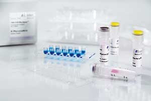
Catalogue Number: K030-0.25 (1 x 250 μl)
Size: 1 x 250 μl (K030-0.25)
HIGHLIGHTS:
- Immediate live observation possible
- Ideal optical properties for transmitted light and fluorescence microscopy
- Optimum cell vitality
- Full serum compatibility
- Visualization of transfection process in living cells
- Efficient, rapid protocol
- Cells can be fixed and dyed
- Compatible with oil immersion
Product Specification
| Application | Proteofection and high-resolution microscopy of living mammalian cells (live cell imaging) |
|---|---|
| Content | PROTEOfectene®, R-Phycoerythrin as positive control, µ-Slides VI (6 channels each) |
| Assays | 90 per kit |
| Shipping | At room temperature |
| Storage | 4°C |
| Shelf life | 1/2 year (after date of delivery) |
| Product Group | Microfection |
The μ-Proteofection Kit VI, developed especially for proteofection, is a unique combination of μ-Slides VI from ibidi GmbH and PROTEOfectene® from Biontex (AFSBio's supplier). The high-performance kit enables living proteofected cells to be observed under high-resolution microscopy.
In the ultra-simple protocol, adherent cells are treated in the μ-Slide with proteoplexes formed from PROTEOfectene® and a corresponding protein, and can subsequently be observed directly in excellent optical quality. Examples of applications include tracking the progress of a fluorescence-tagged protein through the cell from endocytosis to its place of action.
Proteofection of fusion proteins using PROTEOfectene®
PROTEOfectene® is used to introduce the fusion protein into the cell cytosol by means of proteofection. The nuclear localization sequence of the fusion protein is identified by importins and actively transported through the nuclear pores into the nucleus – a process that can be observed using fluorescence microscopy.
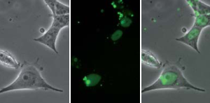
Proteofection of HeLa cells with the protein GST-NLS-GFP and PROTEOfectene®; images clearly show the concentration of fluorescent protein in the cell nucleus adjacent to free proteoplexes and proteoplexes taken up by endocytosis (take-up 20 h after addition of proteoplex).
An example of the efficiency of proteofection is the introduction of the fusion protein GST-NLS-GFP and subsequent nuclear transport of the active protein. It comprises three domains: GST (Glutathion-S-Transferase) is a tag domain serving to purify the protein after expression. NLS (Nuclear Localization Signal) is a short sequence of amino acids which serves to transport the entire protein through the nuclear pores. GFP (Green Fluorescent Protein) finally enables the process to be visualized thanks to its fluorescence in the green band of the electromagnetic spectrum.

Complex of Importin-α and an NLS sequence of a Xenopus frog nucleoplasmin [1]
Literature:
[1] E. Conti, J. Kuriyan; Structure Fold. Des. v8, 329 – 338 (2000) [PubMed ID: 10745017]
Proteofection of Hep G2 cells with R-Phycoerythrin
Proteofection of Hep G2 cells by using the µ-Proteofection Kit VI. Pictures were recorded 24h after addition of the PROTEOfectene/R-Phycoerythrin complex:
 |
 |
|
Picture of cells |
Fluorescence picture: |
Proteofection of HeLa cells with GST-NLS-GFP protein
Proteofection of HeLa cells by using µ-Proteofection Kit VI. Pictures were recorded 24h after addition of the PROTEOfectene/GST-NLS-GFP complex.
 |
 |
|
Picture of cells |
Fluorescence picture: |