Idylle
AgarSqueezer - Agarose-Based Cell Confiner - CRI-SOF
AgarSqueezer - Agarose-Based Cell Confiner - CRI-SOF
Couldn't load pickup availability
The compression height can be chosen among according to the wafer sizes: 2.5µm, 5µm, 30µm, 100µm which can be ordered here.
The device to confine and study your cell behavior within a physiological rigidity range.
Sizes:
- 1 device (CRI-SOF-0)
- 1 device + 1 microscope holder (CRI-SOF-1)
- 2 devices + 1 microscope holder (CRI-SOF-2)
Catalogue Numbers:
- CRI-SOF-0 (1 device)
- CRI-SOF-1 (1 device + 1 microscope holder)
- CRI-SOF-2 (2 devices + 1 microscope holder)
AgarSqueezer is a microscope slide chamber equipped with a molded agar-based compression system. Use it to apply an instant and homogeneous compression on your cells, and study their response to short and long-term confinement.
Physiological rigidity
Contrary to PDMS, the mechanical properties of agarose reproduce stiffness of the in vivo microenvironment (1-150 kPa).
Long-term confinement
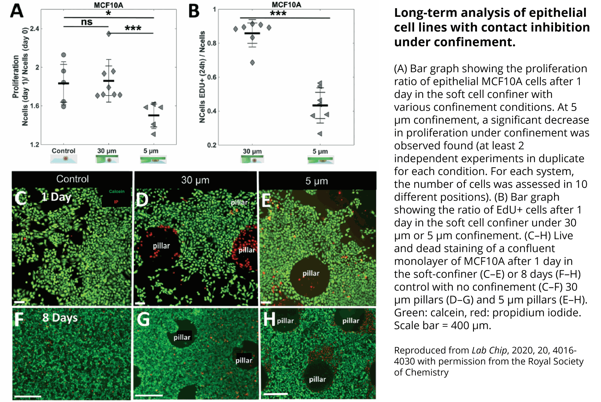
As the porous nature of agarose facilitates nutrient and oxygen diffusion, the AgarSqueezer provides a safe environment for long-term cell culture. Keep your cells confined for up to 10 days!
Flexibility
By modulating confinement level, matrix stiffness & composition or coating with ECM proteins, AgarSqueezer leaves you plenty of options to reproduce environmental cues.
Applications
The AgarSqueezer has been successfully used to confine adherent and non-adherent cells, including:
- Human: primary T-lymphocytes, TF1 & ML2 leukemic cells, HS27A fibroblasts, MCF10A breast cells, MDA-MB-231 breast cancer cells, U-2 OS osteosarcoma cells, PC-3 & DU 145 prostate cancer cells, HT29 & HCT116 colorectal adenocarcinoma cells and HT1080 fibrosarcoma cells
- Murine: megakaryocytes, osteocyte-like cells MLO-Y4, primary muscle cells & primary dendritic cells
- Plant: cells from Arabidopsis roots
- 3D cell cultures: mice gastruloids
Compatible assays:
- Live imaging*: cell viability, proliferation, migration, morphology, nuclear deformability, cytoskeleton reorganization, cell tracking, etc.
- Standard molecular analysis (immunostaining, western blot, qPCR, flow cytometry, etc.): changes in gene/protein expression, proliferation, morphological analyses, etc.
*Cells can be added with fluorescent probes or stimulated with drugs during confinement.
Imaging modes:
Inverted microscopes equipped with a standard adjustable petri dish holder, with any imaging mode: phase-contrast, epifluorescence, confocal, etc.
Kit contents
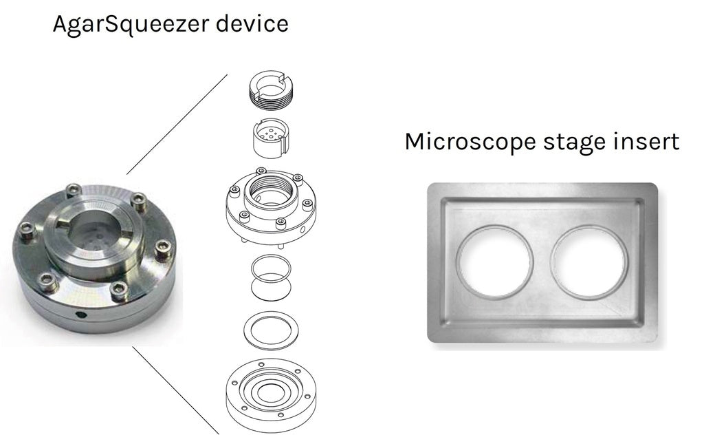
- 1x Agarsqueezer compression device
-
1x 16G flat cut needle to make holes in the agar gel, facilitating diffusion of culture medium or drugs during the experiment
- 1x 20G flat cut needle (same as above)
- (Optional) 1x microscope stage insert (102.5mmx143.5mm), to hold and image up to 2 AgarSqueezers in live imaging chambers (Okolab or equivalent).
In addition to the basic kit contents, it is recommended to use a silicium wafer to mold your confining pillars in agarose. There are four different wafers to mold pillars of various heights. Choose among the four pillar heights available depending on your desired application and add silicium wafer(s) to your order here.
Additional resources:
Results:
Human primary T-lymphocytes migrating in confined conditions
Human T-lymphocytes isolated from blood were seeded in the Agarsqueezer and confined under 5µm pillars. Images were taken every 10sec for 16.5min using an inverted Zeiss Z1 automated microscope (10X objective).
Credits: Marie-Pierre Valignat – Adhesion & inflammation lab, Aix-Marseille University, France
Arabidopsis root cells confined using Agarsqueezer
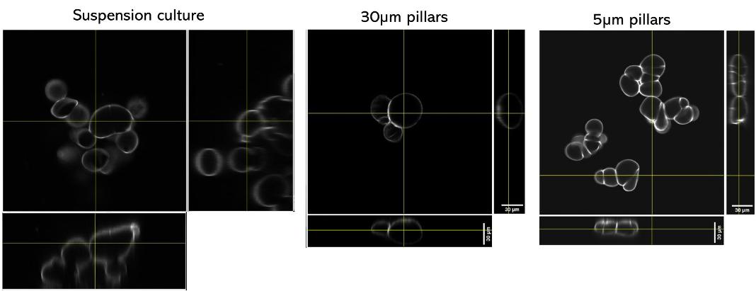
Arabidopsis thaliana Col-0 root cells stained with Calcofluor (cell wall) and imaged with a confocal microscope either in a traditional culture setting (left), or after 24h of confinement in the Agarsqueezer using the 30µm (middle) or 5µm (right) pillars.
Credits: Léa Bogdziewiez - UPSC - Sveriges lantbruksuniversitet, Sweden
Compression of mouse primary myoblasts using AgarSqueezer
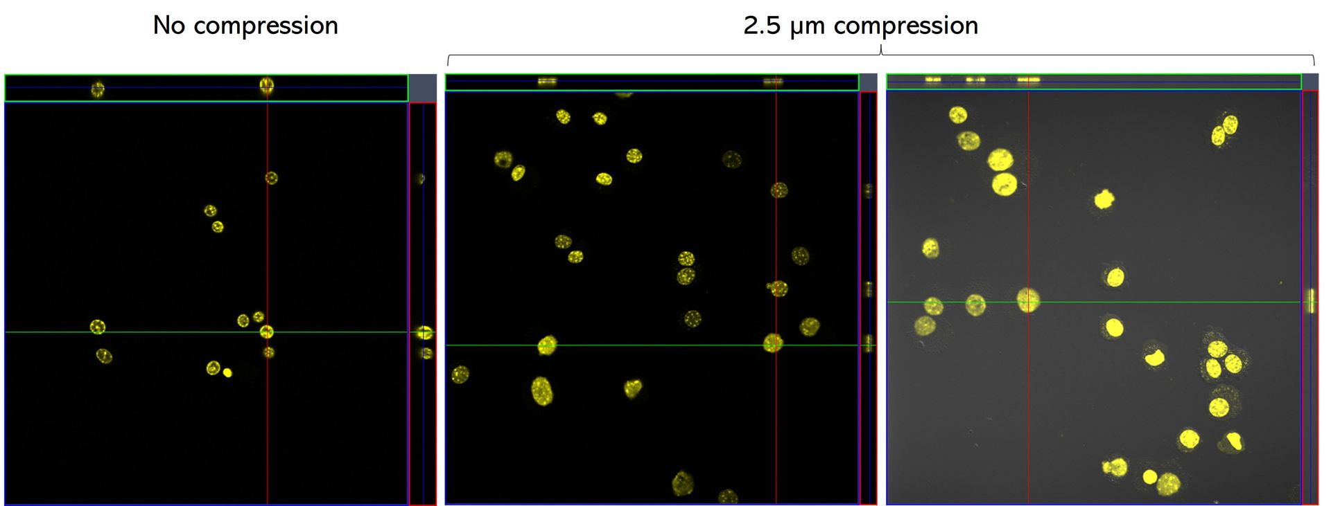
C57 primary myoblasts stained with Hoechst were imaged in the AgarSqueezer before compression (left panel), and after 1.5h of compression under 2.5 µm height pillars (middle & right panels).
Credits: Dr. Hind Zahr & Dr. Alice Varlet, Lammerding Lab – Meinig School of Biomedical Engineering, Cornell University, United States
Observation of vesicles at septum during cell division of mutant E. coli (W3110 tolR - Palmcherry), in LB 1/2 medium, using Chitozen.

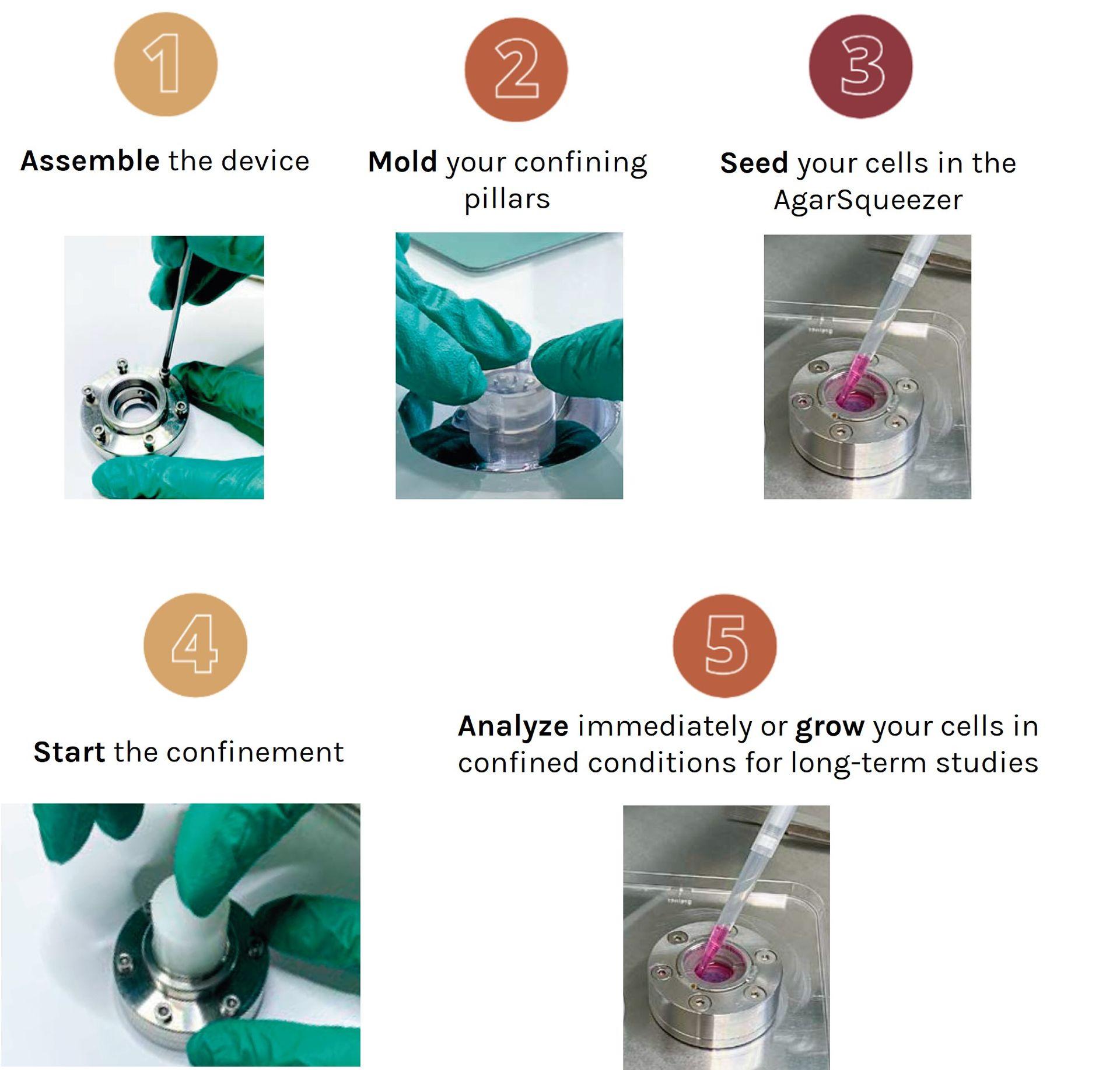
How To Use (In Pictures):