
Idylle
ColorFlux - Visual Indicator of Bacterial Efflux - OSI-STA
ColorFlux - Visual Indicator of Bacterial Efflux - OSI-STA
Couldn't load pickup availability
Fast, reliable and non-toxic solution for measuring bacterial efflux.
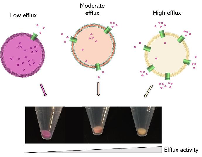
ColorFlux is a fast, reliable and non-toxic solution for assessing bacterial efflux for your research on antibiotic resistance. This coloured fluorescent compound quickly accumulates within bacteria as a function of their efflux pump activity. Simply add ColorFlux to your bacteria and watch them change colour.
How does it work?
ColorFlux staining was shown to reflect the activity of well-characterized efflux pumps from the major facilitator superfamily and ATP-binding cassette families in a variety of Gram+ bacteria:
- NorA, MepA, MepB, PatA, PatB (Staphylococcus aureus & Streptococcus pneumoniae)
- BmrA (Bacillus Subtilis)
The list will be updated regularly according to the feedback of users.
Technical information
Bacterial species compatibility : Gram +
Tested and validated on : Staphylococcus aureus, Bacillus subtilis, Streptococcus pneumoniae.
Colour: Red
Form: liquid
Detection methods: visual & fluorescence. Also compatible with single-cell approaches (live imaging and flow cytometry)
Peak excitation/emission wavelengths: Ex:530nm/Em:650nm
Storage: at 4°C protected from light for 8 months
1 kit contains 1mL of an aqueous solution with a concentration of 1 mg/mL for a final volume of 1L of screening medium
Additional Resources:
Results
ColorFlux staining of MepA&B mutants in Staphylococcus aureus
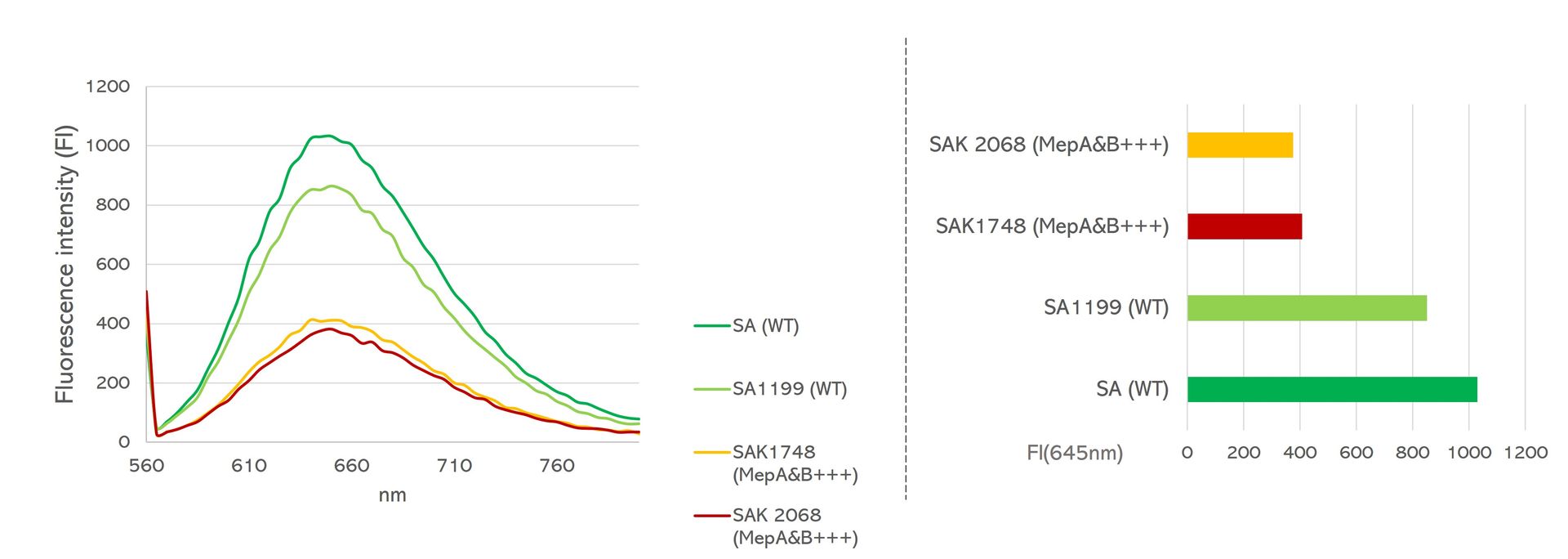
Left graph shows the fluorescence signals detected for each strain using an Ex=530nm, and right graph shows corresponding fluorescence intensity values obtained for each strain at Em=645nm. WT = Wild Type strains, MepA1B+++ = MepA&B overexpressing strains.
Credits: Mrunal Patil & Jean-Michel Bolla, Aix-Marseille Université – 2023
ColorFlux staining of NorA mutants in Staphylococcus aureus
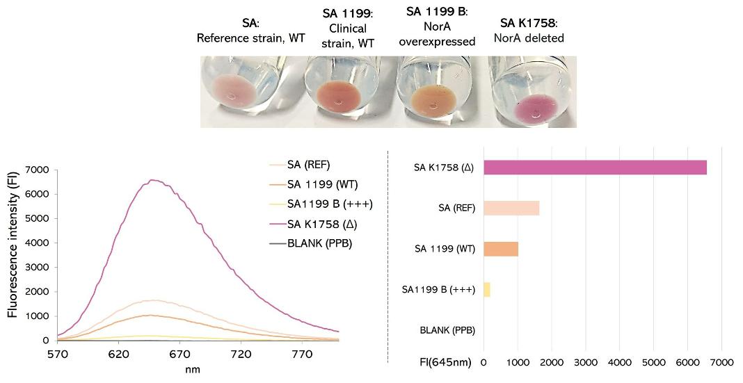
Top picture shows pellet colour variations according to NorA expression. Bottom figures show the fluorescence signals detected for each strain using an Ex=530nm.
Credits: Mrunal Patil & Jean-Michel Bolla, Aix-Marseille Université – 2023
ColorFlux staining of PatA/PatB mutants in Streptococcus pneumoniae
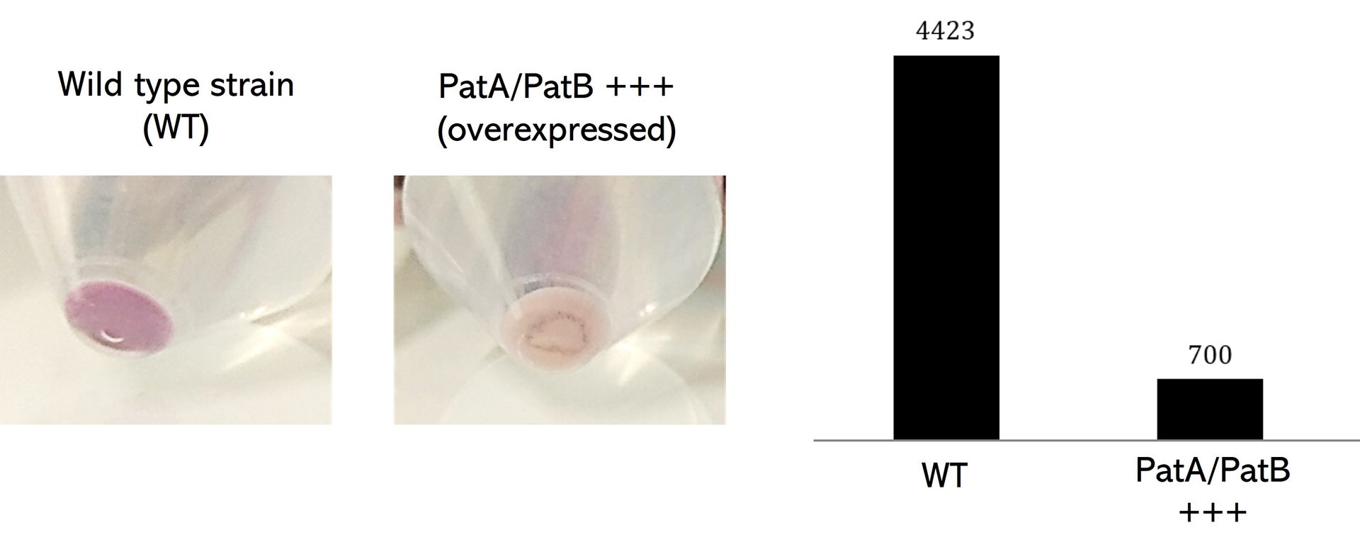
Left pictures show pellet colour variations according to PatA/PatB expression. The right graph show fluorescence signals detected for each strain using an Ex=530nm.
Credits: Mrunal Patil & Jean-Michel Bolla, Aix-Marseille Université – 2023
ColorFlux staining of BmrA mutants in Bacillus Subtilis
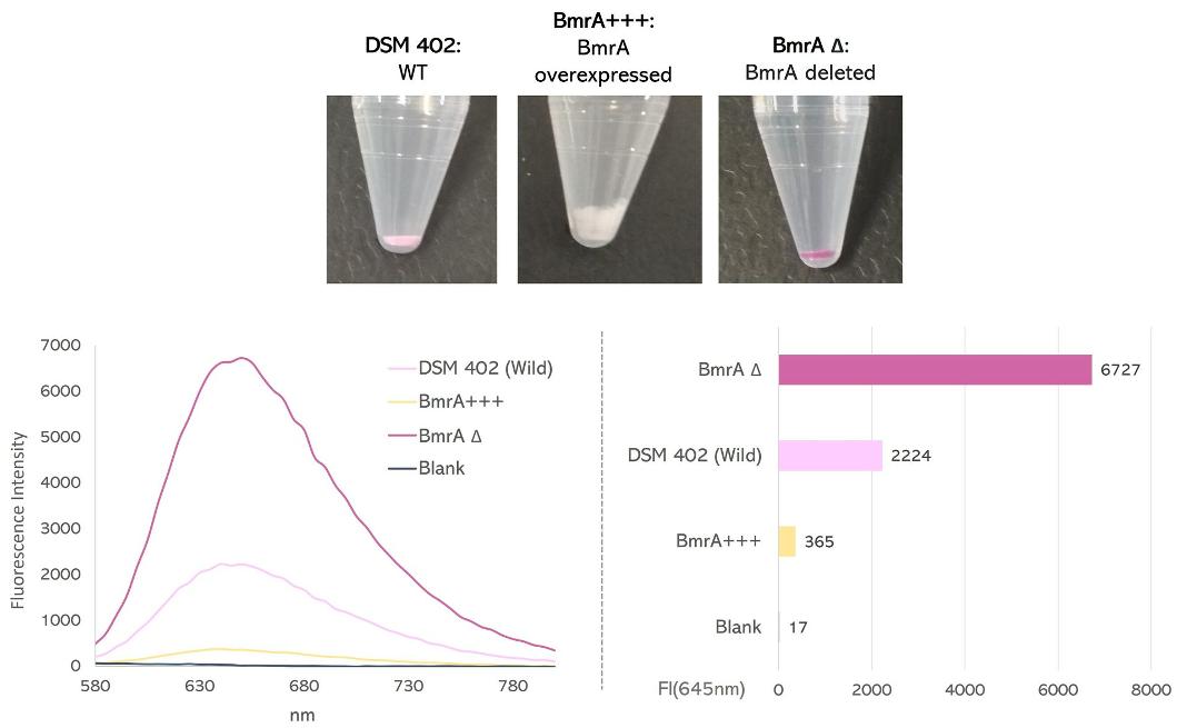
Top picture shows pellet colour variations according to BmrA expression. Bottom figures show the fluorescence signals detected for each strain using an Ex=530nm.
Credits: Mrunal Patil & Jean-Michel Bolla, Aix-Marseille Université – 2023
Bacillus subtilis bacteria (moderate activity) stained with ColorFlux
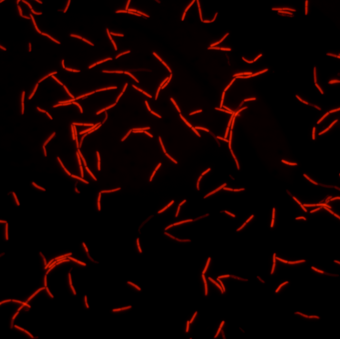 Credits: Mrunal Patil & Jean-Michel Bolla, Aix-Marseille Université – 2023
