


Specific and photostable "Turn-on" fluorescent molecule for high quality imaging of lipid droplets. Kit corresponds to 250mL working solution.
Catalogue Number:
IDY-LIPI-250 (Kit corresponds to 250mL working solution)
Sizes:Key advantages of LipiTrace, a turn-on molecule that makes lipid droplets fluorescent:
- Specificity: Obtain highly specific signal with low noise, resulting in precise image analysis
- Photostability: No risk of photobleaching in your sample, allowing for long-term studies
- Co-labeling: Use the red and far-red channel to label other organelles or proteins
- Universal: Compatible with live staining in mammal cells (lung cancer cell line), yeast, and microalgae.
- Easy to use: Add the solution to your cell media, wait for 5 minutes and start observation (no need to wash).
How does LipiTrace work?
In an acqueous environment (cell cytosol, cell media, Golgi apparatus, mitochondria, endoplasmic reticulum ...), LipiTrace is not fluorescent. However, when it enters inside a lipid droplet, which is apolar, LipiTrace will fluoresce.
This is due to a change of shape depending on polarity: in apolar environment, it adopts a flat shape that enables strong fluorescence. In a polar area, it stays dark due to a twisted shape that blocks fluorescence.This "Turn-ON" property makes it great for imaging, giving high contrast and reduced background signals.

Technical information about LipiTrace
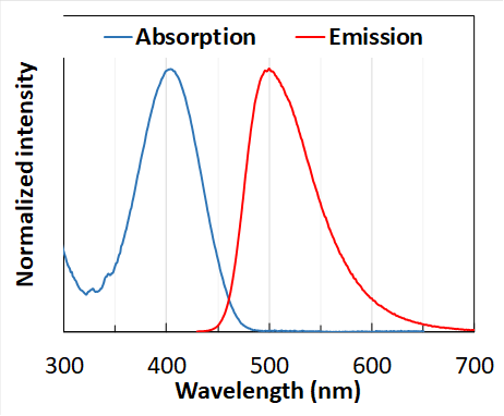
General color of fluorophore: Blue-Green
Excitation wavelength: 360-450nm
Emission wavelength: 460-560nm
Product is sold as a powder, corresponding to 250mL of working solution.
The powder can be stored at room temperature, away from light, for 12 months. Once resuspended in DMSO, the product should be aliquoted and stored at 4°C, away from light. Avoid repetitive opening of the same vial. In these conditions, product will be stable 12 months.
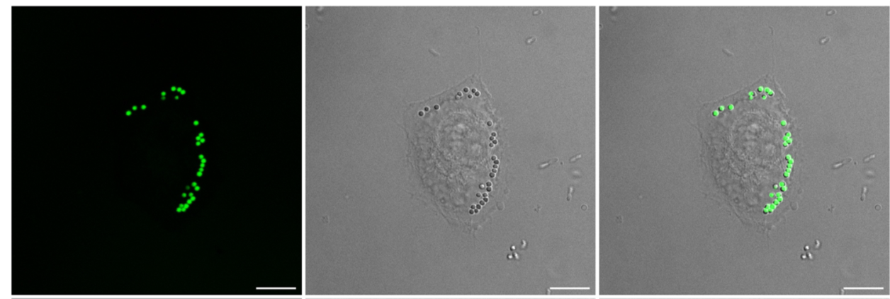
Representative images of lung cancer cells after additIon of LipiTrace to the cell media. (Acquisitions were realised on a confocal microscope equiped with a X63 immersion oil objective, λExc: 440 nm, λEm: 500/50nm, scale bar = 10 μm)
LipiTrace staining is highly specific and has higher signal to noise ratio than BODIPY (1)
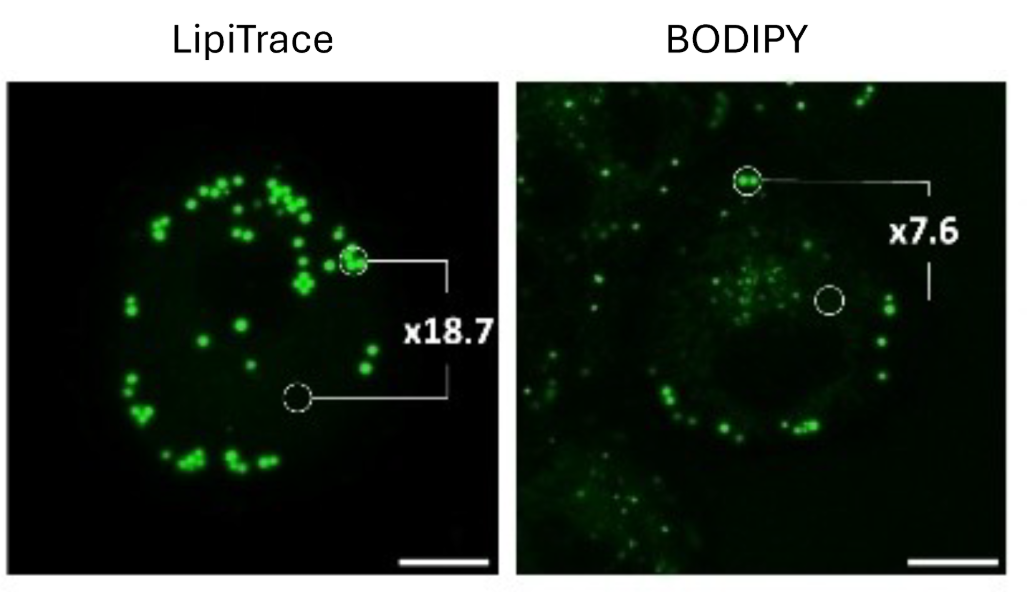 Representative images of lung cancer cell after LipiTrace and BODIPY 493/503 staining. Acquisitions were realised with a confocal microscope equipped with a 63× oil immersion objective. Scale bar : 5µm. Fluorescence intensity measured in cells with average values calculated with n=10 measurements.
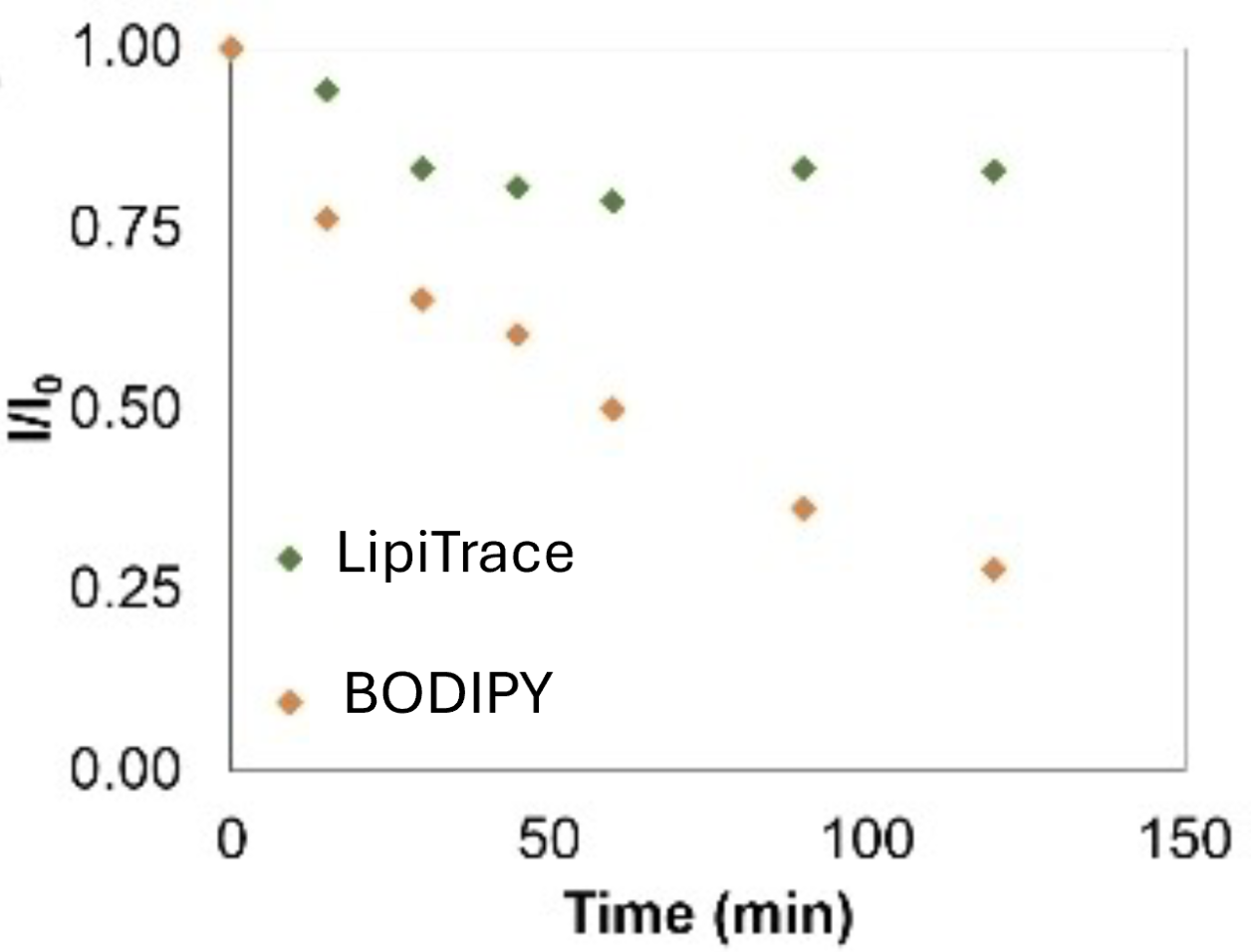
LipiTrace is highly a photostable molecule insuring lack of photo-bleaching, compared with BODIPY (1)
Labeling of live yeast lipid droplets with LipiTrace (2)
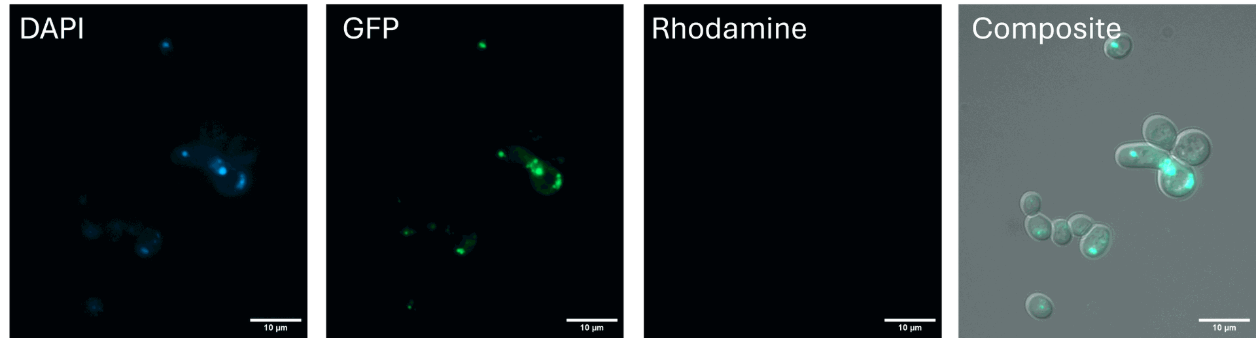 Representative images of the signal obtained in the different channels after addition of LipiTrace to the culture. Acquisitions were realised with an epifluorescence microscope ( Zeiss Axioimager M1). Scale bar =10 µm. © INRAE, Marine Froissard, UMR IJPB
Labeling of live microalgae (Chlamydomonas reinhardtii) lipid droplets with LipiTrace (3)
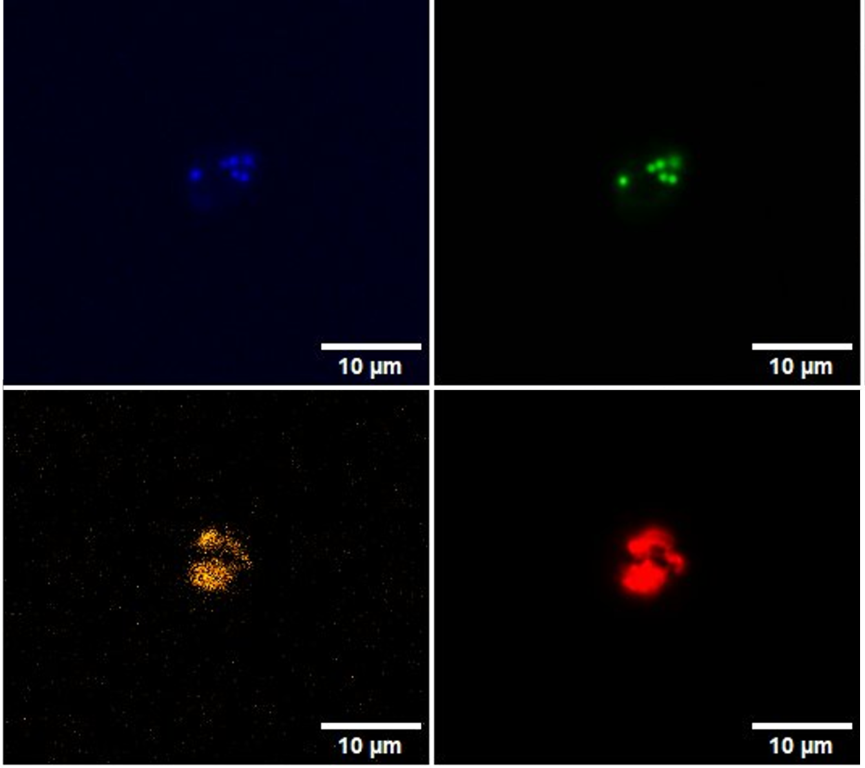 Representative images of the signal obtained in the different channels after addition of LipiTrace to the culture. Microalgae contain chlorophylle, which autofluoresces in the orange/red channel. (Blue : Ex:378/50, Em : 432/36; Green : Ex 474/25, Em: 515/30; Orange: Ex : 554/31, Em: 595/31, Red : 635/16, Em:680/42). Acquisitions were realized with a spinning disk microscope, 40X. Scale bar =10 µm © CEA, Marie Bertrand