

Phimask is a diffraction grating system made for tracking native cellular processes in intact tissues with the following features:
- Compatible with up to 3 colors in the 500-750nm range
- Generating interferences with <20% photon loss
Results
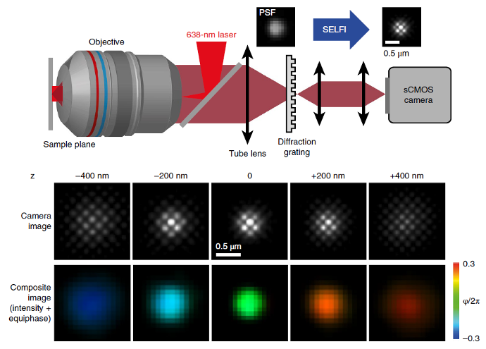
This is a 3D super-resolution design that was published in Bon et al. Nat. Methods 2018. In the image below, you can see that you just have to position the diffraction grating, PhiMask, between the tube lens and the CMOS camera. PhiMask is doing self-interference that change PSF into sub PSF-structuration with very limited photon-loss (less than 20%).
When you have the single-emitter information with sub-PSF structuration, you can simultaneously extract the position in XY and Z.
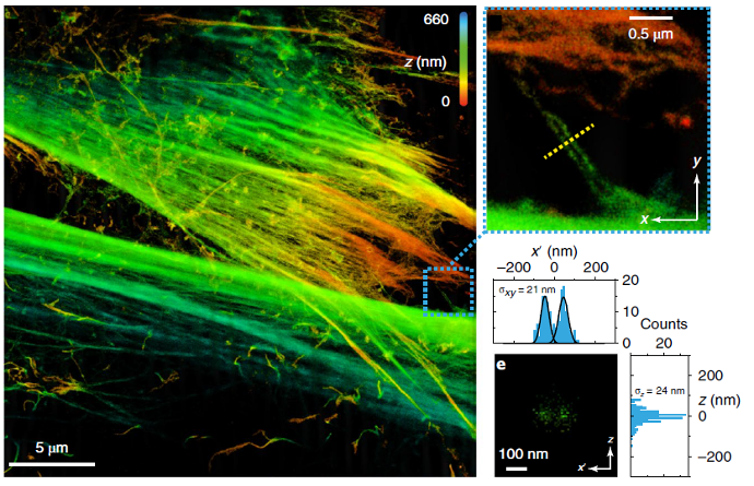
Nanoscale Architecture of Cytoskeletal and Adhesion Networks
Description: Super-resolution imaging reveals aligned filamentous structures with nanoscale spatial organization, highlighting the coordinated arrangement of cytoskeletal elements and associated adhesion components across the cell. The data demonstrate pronounced structural anisotropy and localized clustering at submicron length scales.
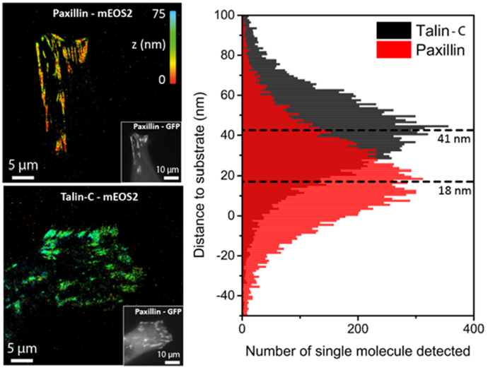
Distinct Axial Localization of Talin and Paxillin in Focal Adhesions
Description: Single-molecule localization analysis shows that talin and paxillin occupy separable vertical layers within focal adhesions. Talin is positioned higher above the substrate than paxillin, indicating a stratified molecular organization consistent with their distinct mechanical and signaling roles.
More information can be found on the Datasheet.