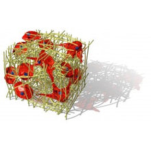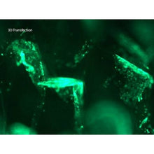
3D-Fect™ is suitable for all kinds of scaffolds and cells. This 3D Transfection Reagent is specifically developed to directly transfect cells cultured in 3D scaffolds. This reagent is based on a novel technology that adds a third dimension to cell culture and transfection. 3D matrices not only add a third dimension to cells’ environment, they also allow creating significant differences in cellular characteristics and behaviours.
Because 3D matrices are routinely used in basic research and therapeutic applications, OZ Biosciences has developed 3D-Fect™ Transfection Reagent specific for 3D-scaffolds. In this way, scaffold-based 3D matrices activated with 3D-Fect™/DNA complexes are colonized by cells which are subsequently transfected in situ and in a more natural environment. This method allows studying tissue engineering and regeneration, tumour invasion, neural differentiation, cellular polarization, tissue formation,colonization, neurite growth...
For Gene Silencing applications, please refer to si3D-Fect
- Highly efficient: cell lines and primary cells
- Specific for 3D Scaffolds
- Compatible with all types of nucleic acids
- Completely biodegradable
- Long term protein expression
- Serum Compatible
Sizes:
- 250 µL (TF20250): Good for 65 to 125 transfections with 1 µg of DNA
- 500 µL (TF20500): Good for 125 to 250 transfections with 1 µg of DNA
- 1000 µL (TF21000): Good for 250 to 500 transfections with 1 µg of DNA
Shipping Conditions: Room temperature
Application
Perfect for all transfections applications in 3D scaffolds such as sponges, matrices, inserts:
- Tissue engineering
- Tissue regeneration
- Tumor invasion
- Neural differentiation
- Cellular polarization
- Tissue formation
- Colonization
- Neurite growth
RECOMMENDED FOR: DNA transfection of cells growing in 3D-scaffolds
Results
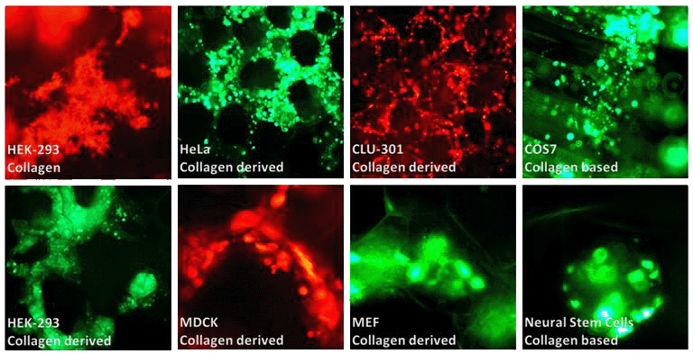
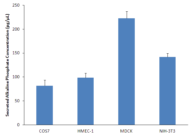
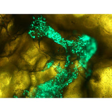
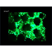
Figure 1: 3D-Fect allows transfection of a variety of cells (cell lines and primary cells) into 3D-scaffolds: Collagen-based and collagen-derived scaffolds were pre-loaded with complexes formed by 3D-Fect™ (ratio 4:1) and 1 μg of either pVectOZ-GFP (green pictures) or RFP plasmids (red pictures). Cells were then seeded and observed under fluorescence microscopy after 48 h.
