
Mold agarose wells to position and image zebrafish larvae and embryos
Stampwell stamps imprint arrays of wells in hydrogels in just one time. Nothing more to do than load your samples into the wells and image. The Stampwell shapes have been optimized for the imaging of fish embryos or larvae up to 20 dpf.
Save time: no need to manually position your samples individually. Fish embryos/larvae will self-position in the wells.
Boost your assay reproducibility: once loaded in the wells, your samples are perfectly positioned, aligned and located on a similar focal plane.
Open system: direct access to embryos/larvae for adding external compounds (i.e. drug screening).
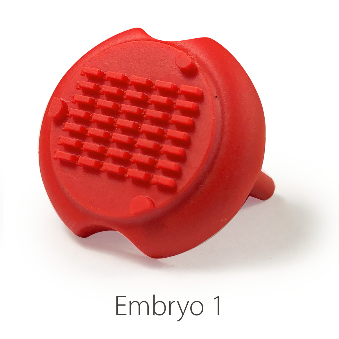
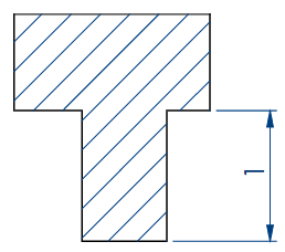
Embryo 1 (i.e. Rectangular)
- Number of pins: 35
- Shape of the pins: rectangular
- Length * width of the wells: 2 mm * 0,65 mm
More information on the Datasheet.
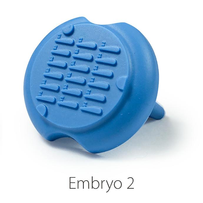
Embryo 2:
- Number of pins: 18
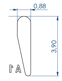
- Shape of the pins: a drop
- Length of the wells: 3.90 mm
- Largest width of the wells: 0.88 mm
More information on the Embryo 2 datasheet.
Larvae 1:
- Number of pins: 5 large pins ideal for the 9-14 dpf zebrafishes + 5 medium pins ideal for the 6-9 dpf zebrafishes + 5 small pins ideal for the 3-6 dpf zebrafishes
- Shape of the pins: fish body and tail
- Length of the 5 large wells: 6 mm, 2.7 for the body and 3.3 for the tail
- Length of the 5 medium wells: 5 mm, 2.25 for the body and 2.75 for the tail
- Length of the 5 small wells: 4 mm, 1.8 for the body and 2.2 for the tail
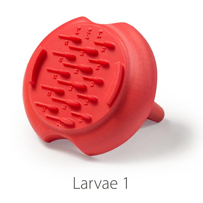
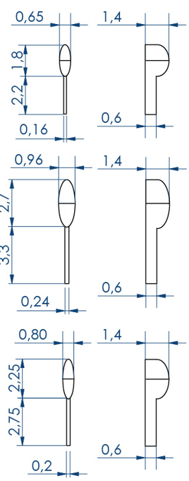
More information on the Larvae 1 datasheet.
Larvae 2:
- Number of pins: 5 large wells ideal for the 15-20 dpf zebrafish or 39-42 dpf medaka larvae + 5 small wells ideal for the 1-2 dpf zebrafish
- Shape of the pins: prism
- Length of the 5 large wells: 10 mm
- Length of the 5 small wells: 3 mm
- Well depth: 1 mm
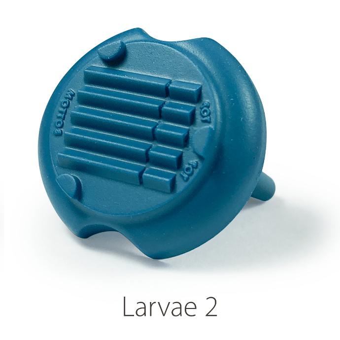
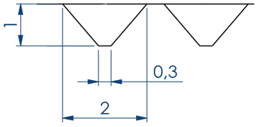
More information on the Larvae 2 datasheet.
Additional resources:
Product overview
Datasheet
Embryo 2 datasheet
Larvae 1 datasheet
Larvae 2 datasheet
Stampwell FAQs
Results:
Parallelized imaging of zebrafish embryos using the Embryo 1 Stampwell 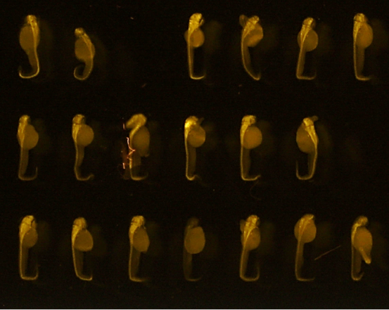
48hpf embryos (anti-HuC/D, ab210554, abcam)
Credits: Matthieu Simion, CNRS, 2022
Improved positioning of 2dpf zebrafish larvae using the Embryo 2 Stampwell
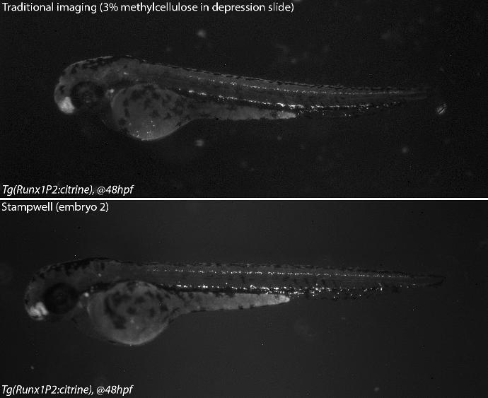 Credits: Dr Rui Monteiro - Institute of Cancer and Genomic Sciences, University of Birmingham, United Kingdom
Zebrafish embryos imaging in agarose wells made with Stampwell Embryo 1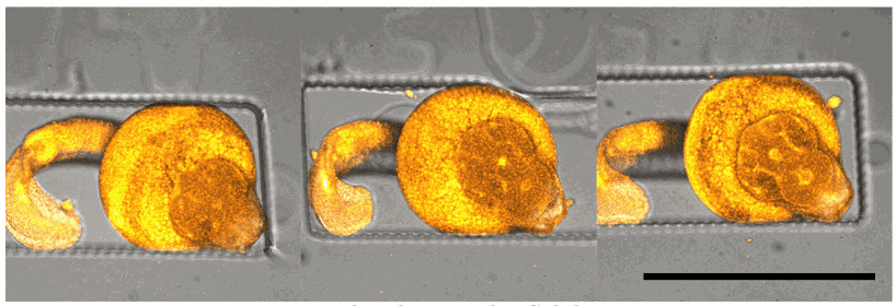
Bar: 1mm (projection of Z-stack - confocal microscope)
Credits: Gaëlle Recher - Bordeaux 2019
Heart imaging in a Larvae 1 Stampwell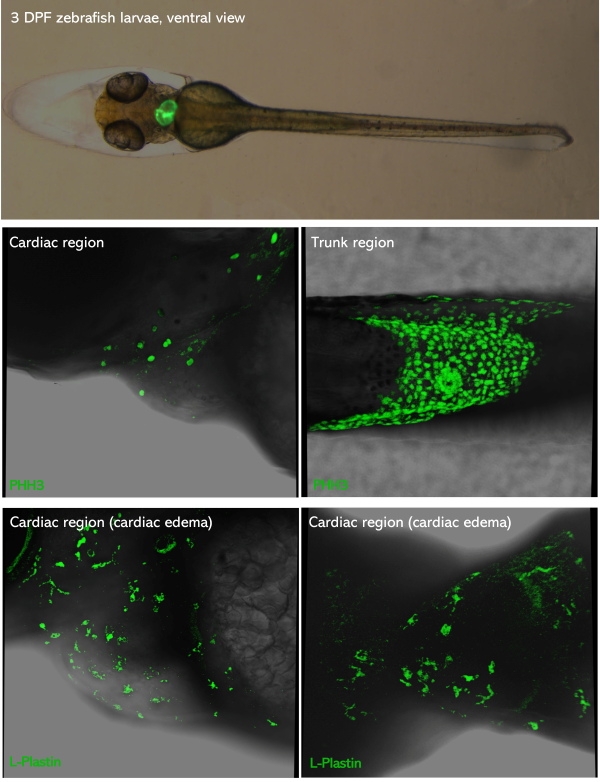
Top picture: green-heart 3dpf zebrafish larvae placed in ventral view. Middle & bottom pictures: 3dpf zebrafish larvae PFA-fixed,
mounted in glycerol and inserted in Larvae 1 Stampwell (anterior to the left). Pictures were taken using a Leica Spectral Confocal SP5.
Credits: Dr. Giovanni Risato, Prof. Natascia Tiso and Alina Ramazanova - Department of Biology, University of Padova, Italy


