


Clearing kit for biological tissues
Kit contents:
- 1x Depigmentation Mix
- 1x Clearing Base
- 1x Imaging Solution
Catalogue Numbers:
- IDY-UBI-S (Depigmentation buffer (20mL), Mix 1 (5mL), Mix 2 (40mL))
- IDY-UBI-L (Depigmentation buffer (200mL), Mix 1 (50mL), Mix 2 (400mL))
- IDY-UBI-MIX2-KIT (Mix 2 (400mL))
Sizes:
- Depigmentation buffer (20mL), Mix 1 (5mL), Mix 2 (40mL) - IDY-UBI-S
- Depigmentation buffer (200mL), Mix 1 (50mL), Mix 2 (400mL) - IDY-UBI-L
- Mix 2 (400mL) - IDY-UBI-MIX2-KIT
UbiClear is an innovative method for optical clearing of biological samples. Thanks to its unique formulation, the UbiClear solution makes thick samples transparent in less than 20 minutes without any toxic compounds. Place your sample in the clearing buffer, incubate at 37°C and transfer it to the imaging solution.
-
Non-toxic
- Formulated without any harsh chemicals, UbiClear can be handled safely and does not deteriorate microscope objectives
-
Compatible with fluorescence labeling
- UbiClear preserves endogenous fluorescence and can be combined with immunostaining protocols
-
Ultra-fast
- Clear your samples within minutes thanks to a 1-step clearing method
-
Versatile
- Clears a vast panel of soft and hard mammalian tissues (bone, teeth), insects and plants.
How does UbiClear work?
UbiClear is an aqueous solution that modifies the optical properties of complex biological tissues by removing lipids and homogenizing refractive indexes. This renders opaque tissues transparent while keeping their original structure intact. As a result, light passes through specimens without any scattering or absorption, yielding high-quality images of entire 3D structures. The UbiClear workflow is straight-forward, and does not require any specific expertise or access to sophisticated equipment.
-
Depigmentation (1h)
- Remove sample coloration by immersing it into UbiClear Depigmentation Buffer for 1h at 37°C.
-
Clearing (20min - overnight)
- Incubate your sample into the UbiClear clearing solution at 37°C until fully transparent. Maximal transparency is achieved in a few minutes for most tissue fragments (<20 min for mouse cerebral cortex and small intestine), and may take up to a few hours for entire organisms.
-
Imaging
- Transfer your cleared sample to the imaging solution (RI = 1.47) for imaging and maintaining transparency on the long-term.
-
Repeat!
- If needed, samples can be re-opacified simply by transferring them to 0.2 M Tris buffer.
UbiClear applications
Sample types: UbiClear can be used to clear a wide spectrum of samples ranging from small 3D cell structures up to entire organisms. So far, UbiClear has shown efficient clearing of:
- 3D cell structures: human embryoid bodies, human mini-brains
- Human tissues: frontal cortex
- Mice tissue sections (up to 1 mm thick): spinal cord, brain (cerebellum, choroid plexus), testes, seminal vesicle, ear (elastic cartilage and hair), lungs, heart, intestine, kidney, skeletal muscle, yolk sac membranes
- Ox tissues (up to 1 mm thick): knee (joint cartilage and bone)*
- Plants: ivy leaves, carrot slices, beet roots, microcallus & callus organoids, nasturtium stamen, melon seeds, sprouted tomato seeds, rose stem slices
- Whole organs: mouse brain, heart, intestine, eye, teeth*, dorsal root ganglia
- Whole organisms: Colisa Lalia, adult zebrafish, firebugs (Pyrrhocoris apterus), wasps, grasshoppers, shield bugs (Graphosoma lineatum), flies, cockroaches, spiders
- Crustaceans: woodlouse
- Molluscs: slug, mussel (including shell)*
*for these samples, an additional step of demineralization needs to be performed before clearing.
Compatible assays: label-free anatomical studies (autofluorescence, volumetric imaging), dye-based assays (Evans blue, albumin-FITC), immunostaining. UbiClear preserves endogenous fluorescence.
Microscopy techniques: confocal, spinning-disk, light-sheet microscopes†.
†Extra diluting buffer can be purchased separately to make more imaging solution if needed (i.e. for use with light sheet microscopes).
Additional resources
- Product overview
- Safety Data Sheet - Depigmentation buffer
- Safety Data Sheet - Mix 1
- Safety Data Sheet - Mix 2
Results
Fast and versatile clearing of diverse animal tissues using UbiClear
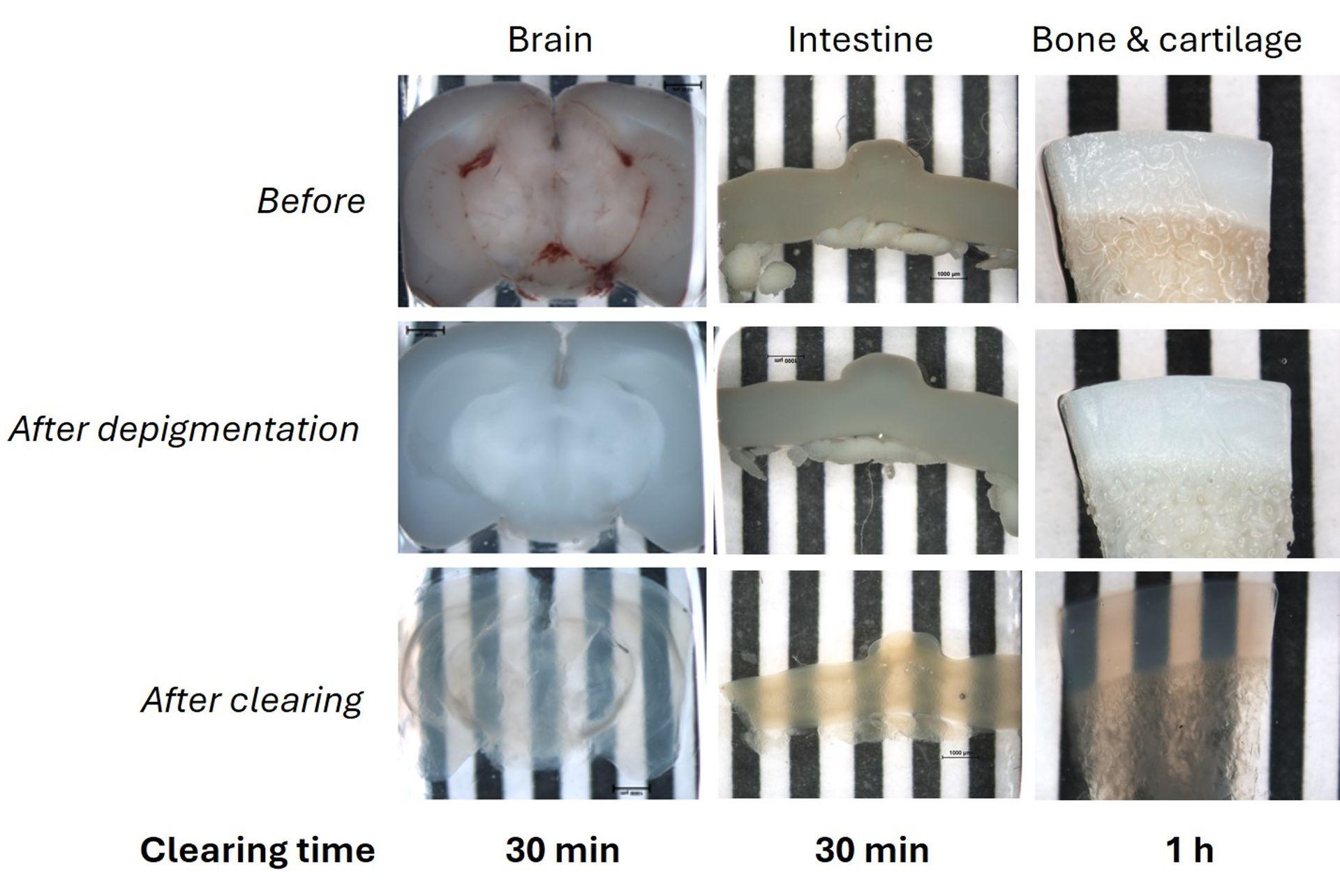 1 mm thick mice brain slices (fat tissue), 2 cm mice intestine fragments and a 1 mm thick ox knee slices (joint cartilage and bone, calcified tissue) were cleared using the UbiClear method and imaged on a striped target. Ox knee joint samples were decalcified and all samples depigmented and cleared using UbiClear. The target stripes are visible through the sample after clearing for less than an hour.
 Credits: Brigitte Delhomme & Martin Oheim (CNRS, SPPIN UMR8003)
Visualizing Glial fibrillary acidic protein (GFAP) expression in mouse brain
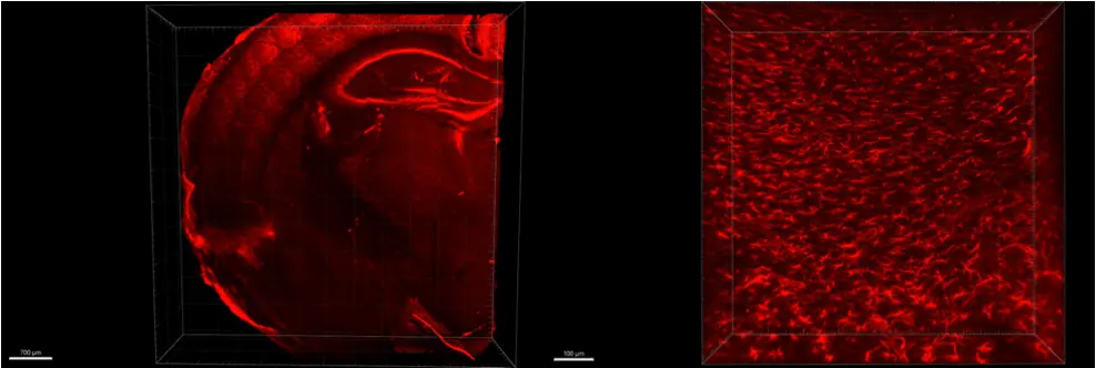 Indirect immunofluorescence and clearing was performed on 1 mm thick sections of formaldehyde-fixed mouse brain using GFAP antibody and secondary antibody Alexa 555 fluorophore. On the left the whole slice was imaged in mosaic with 10XNa0.3 objective, on the right 40XNa1.0 was imaged in the cortex of the mouse brain.
Credits: Diane Derrien, Brigitte Delhomme & Martin Oheim (CNRS, SPPIN UMR8003)
Cleared mouse dorsal root ganglia combined with immunohistochemistry
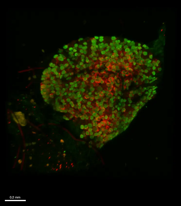 Immunofluorescence and clearing was performed on whole fixed dorsal root ganglia from a transgenic mouse model that expresses both (i) a genetically-encoded GCaMP6 calcium sensor in sensory neurons as well as (ii) a hematoglutinin-tagged chemogenetic receptor in satellite glial cells. An anti- GFP (green) and an anti-hematoglutinin (red) antibodies were used to amplify GCaMP6 staining and reveal satellite glial cells, respectively. The whole ganglia was imaged using a confocal microscope Axio LSM710 (Zeiss) and 10XNa0.3 WD 5mm air objective, in 620 µm depth and z 2 µm. 3D reconstruction was obtained with Imaris viewver software (oxford instruments).
 Credits: Andreia Ramos (Université Paris Cité, CNRS, INCC UMR8002, Cendra Agulhon’s Group) and , Brigitte .Delhomme (Université Paris Cité, CNRS, SPPIN UMR8003, Martin Oheim’s laboratory). Courtesy of Cendra Agulhon.
Mapping Iba1 expression in a cleared mouse spinal cord
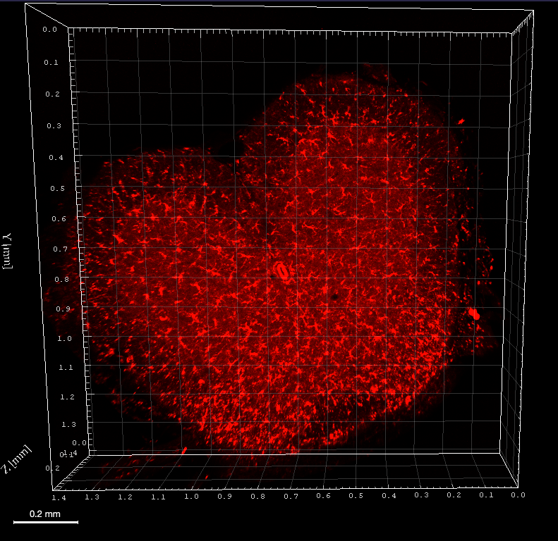 Immunofluorescence was performed on a 500µm mouse spinal cord section after depigmentation. Iba1 antibody, a rabbit polyclonal antibody, and Cy3 secondary antibody were used for incubations. Iba1 was imaged using an Axio LSM710 (Zeiss) confocal microscope with 10XNa0.3 WD 5mm air objective, zoom 0.6 in 270µm depth and z 2µm. 3D reconstruction was performed using Imaris viewver software (Oxford Instruments).
Credit : Clearing B.Delhomme & M.Oheim (CNRS, SPPIN UMR8003)
Insect clearing using UbiClear
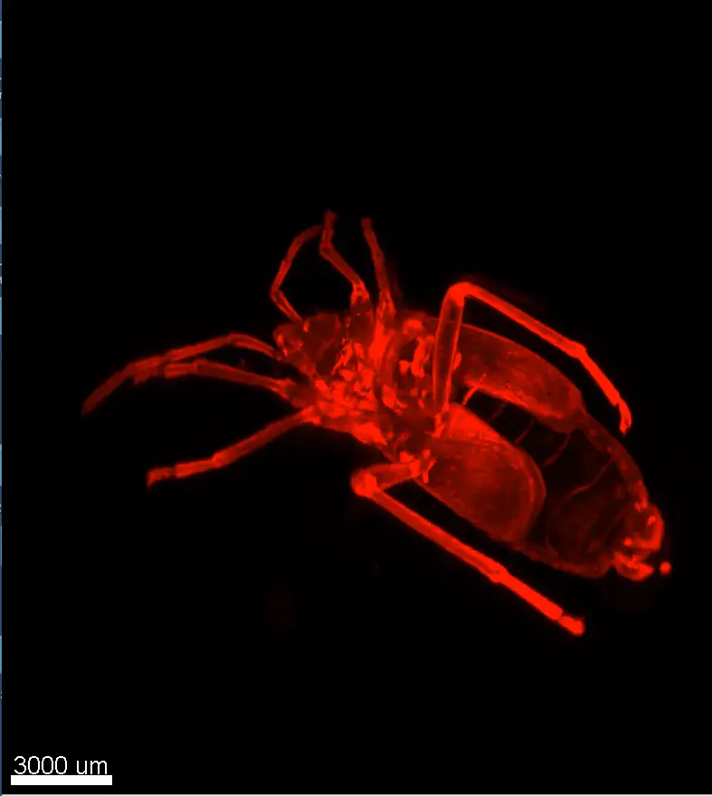 A firebug (Pyrrhocoris apterus) was depigmented, embedded in low melting agarose and cleared using UbiClear. The image showing autofluorescence signal at 488 nm was acquired using a light-sheet ultramicroscope (Labvision). 2,400 images were stitched for the 3D reconstruction.
 Credits: Clearing: Brigitte Delhomme (CNRS, SPPIN UMR 8003), Imaging: J. Fernandes (Institut Pasteur, Paris) & Martin Oheim (CNRS, SPPIN UMR8003)
Whole brain clearing using UbiClear
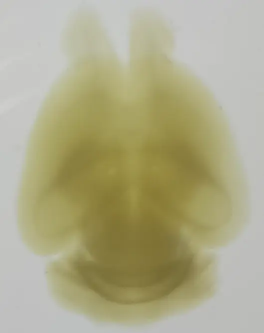 A whole brain from a 4-month old mouse was depigmented and cleared using UbiClear. The picture was taken 48h after clearing.
 Credits: Brigitte Delhomme & Marwa Moulzir (CNRS, SPPIN UMR 8003)
One-step clearing of the small intestine
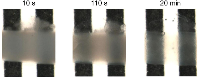 A 1.5 cm long mice intestinal fragment was cleared using UbiClear and placed in a home-made tank with a black-and-white stripe pattern. Images were acquired on a SMZ800 stereo macroscope (Nikon, Amstelveen, The Netherlands) after 10sec, 110sec and 20min of incubation in the UbiClear clearing solution. Michelson’s contrast calculation revealed that 71% transparency was reached in less than 15 min.
 Source publication: Hazart D. et al, 2023
Imaging deep blood vessel network in mouse cerebellum
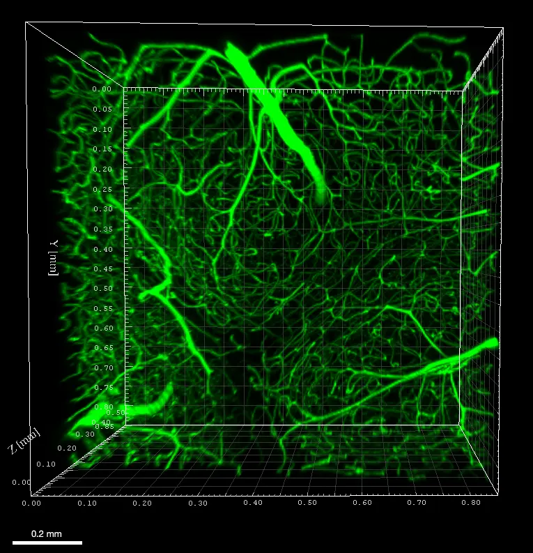 Mouse was perfused with FITC albumin and sacrificed to image blood vessels in the cerebellum. A 500µm tissue slice was cleared without depigmentation and imaged using an Axio LSM710 (Zeiss) confocal microscope with 488nm laser and 10XNa0.3 WD 5mm air objective, in 508µm depth and z 4µm. 3D reconstruction was performed using Imaris viewer software (Oxford Instruments).
Credits : Mouse painting vessels X.Bai (University of Saarland, Homburg (DE)), Clearing B.Delhomme (CNRS, SPPIN UMR8003, Paris (Fr)) , F.Licata (UMS Paris-cité University (Fr) & Martin Oheim (CNRS, SPPIN UMR8003, Paris (Fr))
Detailed morphological characterization of the enteric nervous system
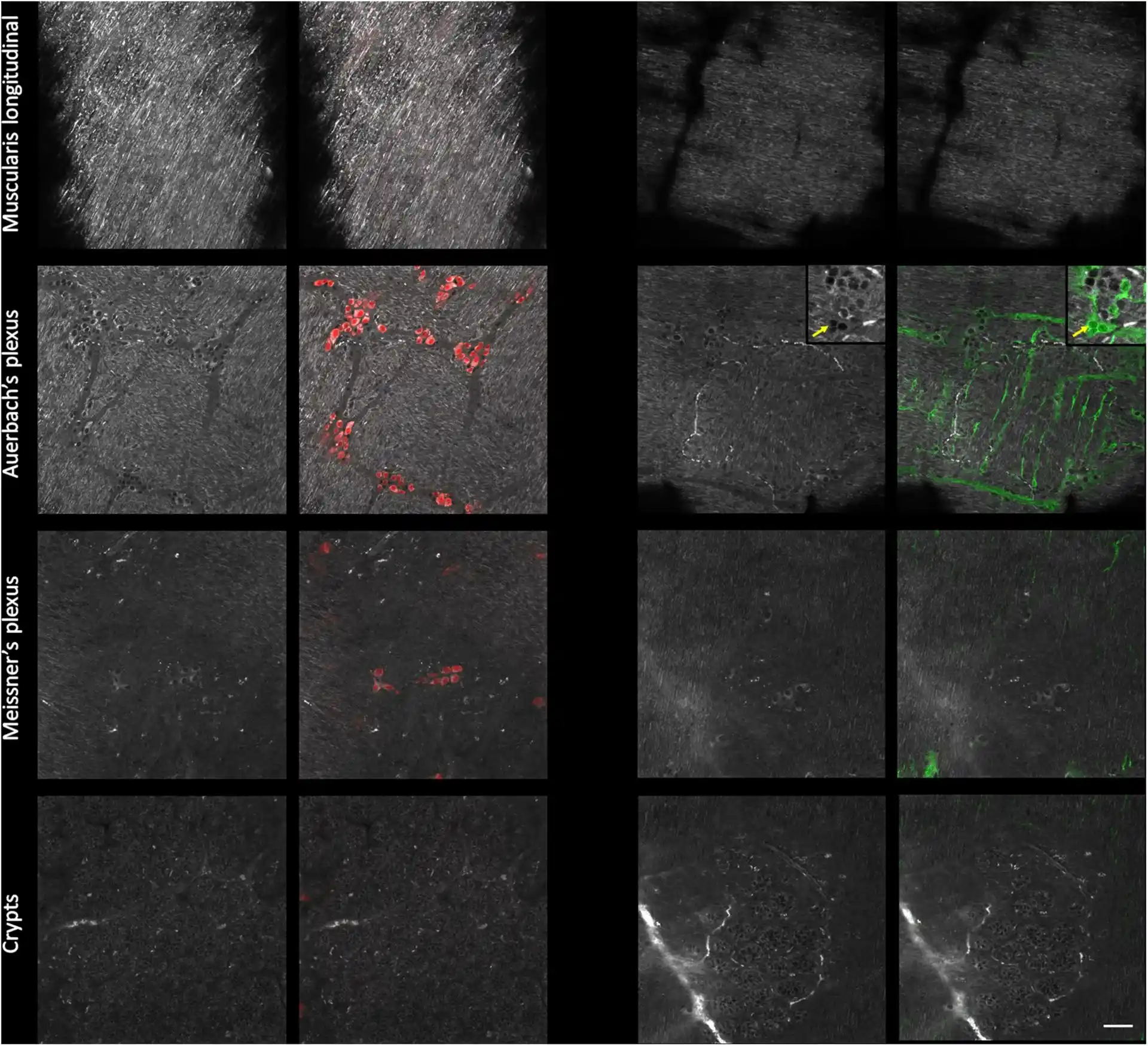 Mice small intestine fragments were cleared using UbiClear and immunolabeled against a pan-neuronal marker (red) and an enteric-glia marker (green). Single autofluorescent images (grayscale) and fluorescence images were taken at different depths in the intestinal wall (muscularis longitudinalis, Auerbach’s plexus, Meissner’s plexus and crypts). Images were acquired on a LSM880 inverted confocal Airyscan microscope (Zeiss).
 Source publication: Hazart D. et al, 2023