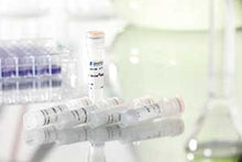
High-efficiency antibody proteofection reagent for mammalian cells
Catalogue Numbers:
- E020-0.1 (1 x 100 μl)
- E020-0.25 (1 x 250 μl)
Sizes:
- 1 x 100 μl (E020-0.1)
- 1 x 250 μl (E020-0.25)
HIGHLIGHTS:
- High efficiency and broad range of cell types
- Mono- and polyclonal antibodies of all isotypes
- Ultra-low toxicity – cell vitality is maintained
- Full serum compatibility
- Efficient, rapid protocol
Product Specification
| Application | Proteofection of eukaryotic cells with anti bodies |
|---|---|
| Content | PROTEOfectene® AB, 100 µl IgG (FITC labeled; 100 µg/ml) as positive control |
| Assays | Approx. 100 (24-well) or approx. 20 (6-well) per 100 µl reagent |
| Shipping | At room temperature |
| Storage | 4°C PROTEOfectene®AB; ≤ –15°C IgG |
| Shelf life | 1/2 year (after date of delivery) |
| Product Group | Proteofection |
PROTEOfectene® AB complexates antibodies noncovalently by means of electrostatic and hydrophobic interactions, forming a proteoplex which is taken up by cells through endocytosis and finally releases the antibodies in the cell cytosol while retaining full functionality and maintaining epitopic binding affinity. PROTEOfectene® AB can be used to introduce mono- and polyclonal antibodies of all isotypes.
Used with the incredible range of antibodies on the market, PROTEOfectene® AB creates new possibilities for research and manipulation of cell processes. For example, fluorescence-marked antibodies can be used for immunostaining in living cells, enabling live cell imaging of processes to be taken. By using antibodies against a variety of cell constituents – proteins/enzymes, transmitter substances, educts and products of metabolism – antibody proteofection becomes a method offering many possibilities.

Proteofection of A549 cells with PROTEOfectene® AB and AlexaFluor®488 labeled antibodies directed against the nuclear pore complex. Fluorescent microscopy clearly shows the selective binding of antibodies to nuclear pores.

Proteofection of A549 cells with AlexaFluor®488 labeled antibodies against giantin – a membrane protein of the Golgi apparatus – using PROTEOfectene® AB. Fluorescent microscopy clearly shows the selective binding of antibodies in the region of the Golgi apparatus.


