si3D-FectIN™ is a 3D transfection reagent specifically designed and developed for silencing gene expression in cells cultured in gels (or hydrogels).
3D matrices not only add a third dimension to the cells’ environment, they also allow creating significant differences in cellular phenotype and behaviour. In this way, 3D matrices bearing complexes formed with si3D-FectIN™ reagent and siRNA are colonized by cells to be transfected in a more natural environment.
For DNA Transfection, please refer to 3D-FectIN
- Highly efficient for gene silencing in 3D matrices
- Ideal for any gel and hydrogel
- Dedicated to short nucleic acid sequences (siRNA, miRNA…)
- Long term gene silencing
- Universal (primary cells and cell lines)
- Serum Compatible
Sizes:
- 250 µL (STN50250): Good for 30-65 transfections with 50 nM siRNA in 50 µL Gel
- 500 µL (STN50500): Good for 65-125 transfections with 50 nM siRNA in 50 µL Gel
- 1 mL (STN51000): Good for 125-250 transfections with 50 nM siRNA in 50 µL Gel
Storage: +4°C
Shipping Conditions: Room temperature
Application
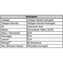
Perfect for all transfection applications in 3D hydrogels such as collagen, hyaluronic acid, PEG, fibrin, laminin:
- angiogenesis
- tube and acini formation
- colonization
- neurite growth
- tissue engineering
- tissue regeneration
- tumour invasion
- neural differentiation
- cellular polarization
- tissue formation…
RECOMMENDED FOR: Gene silencing of cells growing in 3D-hydrogels.
Results
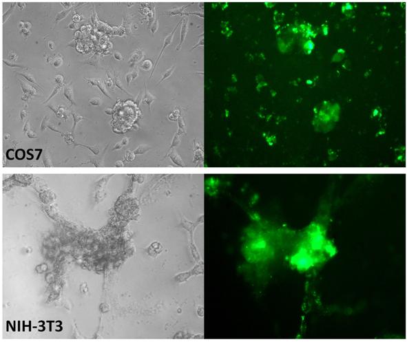
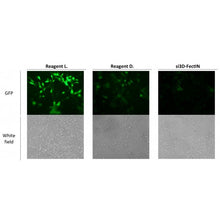
Figure 1: si3D-FectIN™ allows efficient siRNA transfection in hydrogels: Collagen-derived hydrogels were loaded with 50 nM of fluorescently labelled siRNA complexed to 4 µL of si3D-FectIN™ transfection reagent. Transfection efficiency of COS-7 and NIH-3T3 cells was observed after 48 h by fluorescence microscopy.
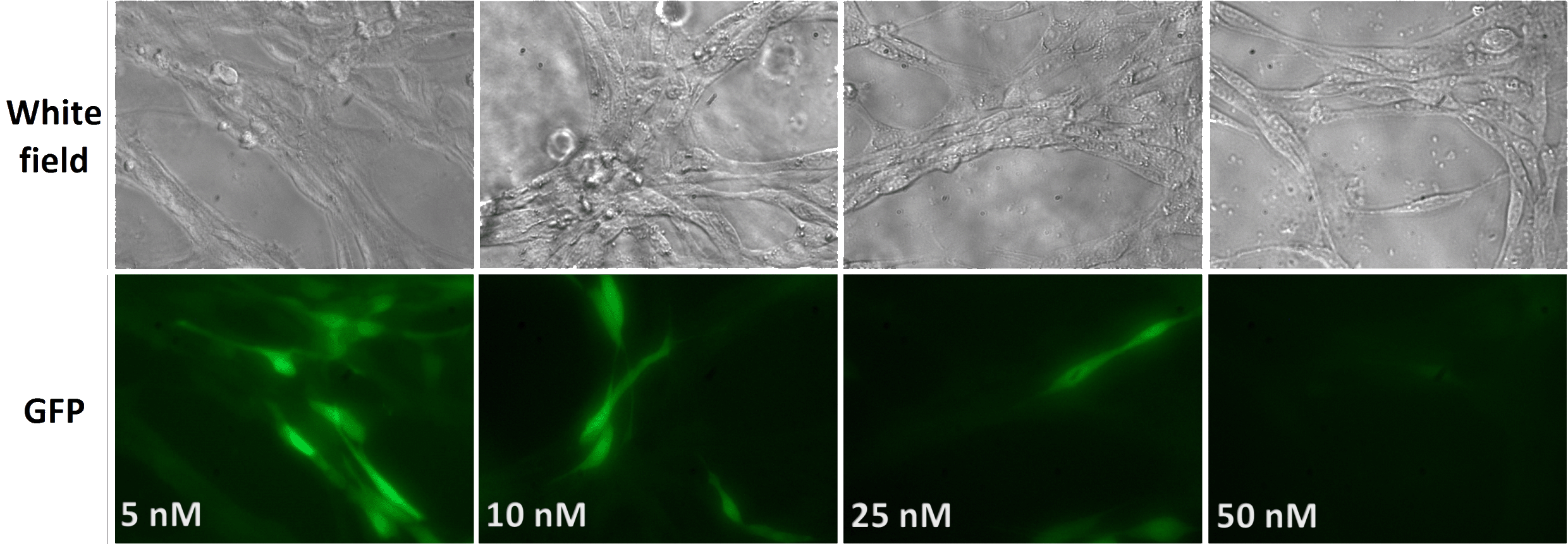
Figure 2: si3D-FectIN™allows optimal gene silencing in 3D-hydrogels: Collagen-derivedhydrogels were loaded with complexes formed by 4 µL si3D-FectIN™ and several concentrations of siRNA (5-50mM) directed against GFP. NIH-3T3-GFP were added and gene silencing was observed after 72 h by fluorescence microscopy.


