NeuroMag Transfection Reagent is the first dedicated Magnetofection™ transfection reagent for neurons from 1 DIV to 21 DIV. It has proven to be extremely efficient in transfecting a large variety of primary neurons such as cortical, hippocampal, dorsal root ganglion and motor neurons with all types of nucleic acids. Moreover, high transfection efficiency was also achieved in primary astrocytes, oligodendrocyte precursors or neural stem cells as well as other cell lines (C6, B65, PC12, N2A...). Due to its unique properties, NeuroMag allows following transfected neurons during several days.
- Great efficiency, ideal for primary neurons
- Efficient from 1 DIV to 21 DIV
- Non toxic and completely biodegradable: high transfected neurons viability
- Ready-to-use, straightforward and rapid
- For all types of nucleic acids
Sizes:
- 200 µL (NM50200): up to 65 transfections with 1µg of DNA
- 500 µL (NM50500): up to 165 transfections with 1µg of DNA
- 1 mL (NM51000): up to 330 transfections with 1µg of DNA
- #KC30800: Starting Kit with Super Magnetic plate + 200 µL of NeuroMag
Storage: -20°C
Shipping Conditions: Room temperature
- This reagent needs to be used with a magnetic plate
Application
NeuroMag transfection reagent:
- Transfection of all types of nucleic acids: DNA, shRNA, siRNA, mRNA, oligonucleotides
- Suitable for all kinds of neural cells:
- Primary neurons: hippocampal, cortical, cerebellar granules, motorneurons
- Neural Stem Cells, embryonic DRG
- Neuronal cell lines: A172, B65, C6, KS-1, N2A, PC12, SH-SY5Y, SKN-BE2, T98G, U251, U87, YH-13...
RECOMMENDED FOR: Transfection of neuronal cells
Results

Figure 1: Rat primary hippocampal neurons efficiently transfected with NeuroMag. Representative image of rat primary hippocampal neurons transfected with 1 μg pVectOZ-GFP and 3.5 μL NeuroMag in 24-well plate. Photos were taken under fluorescence 48H after transfection

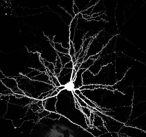
Figure 2: Mouse cortical neuron expressing GFP (3 weeks in culture, 2-3 days after after magnetofection). Image acquisition: Nikon Ti-E microscope equipped with the A1 laser scanning confocal system and a 60x Apo TIRF objectif lens. Results were kindly provided by Dr. C. Charrier (Charrier C. et al., 2012, Cell, Vol 149, Issue 4, 923-935).


